Chip implant restores some vision lost to retinal disease
A study’s favorable outcome gives hope to a UW Medicine surgeon who treats patients’ degenerative eye conditions.Media Contact: Brian Donohue - 206-457-9182, bdonohue@uw.edu
A wireless microchip implanted in the back of patients’ eyes restored some vision for a group of people suffering from an incurable, progressive retinal disease, according to a study published Oct. 20 in the New England Journal of Medicine.
Currently the implant is an investigational device only, but if approved by public health agencies, “not only could this benefit patients with macular degeneration, but also other retinal degenerations like, for example, Stargardt disease (and) retinitis pigmentosa,” said Dr. Lisa Olmos de Koo, who helped design the study. She is a professor of ophthalmology at the University of Washington School of Medicine and a surgeon who cares for patients with debilitating retinal diseases.
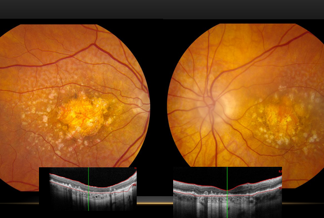
The trial involved 38 patients suffering from geographic atrophy, a type of age-related macular degeneration. In this condition, cells at the center of the retina die, resulting in a black spot at the center of their vision. About 5 million people globally are estimated to have this condition.
“For patients, that impacts them severely because they're unable to read, see faces, tell time, and that can really impact independence for older adults,” Olmos de Koo said, adding that the condition typically emerges among people age 55 or older. The only current medical treatments are monthly injections into the eye, which slow the condition but do not reverse it.
The trial participants were implanted with the chip between 2021 and 2022 at 17 European sites.
“The implant was placed under their retina, and then we collected data. We looked at how the patients functioned with and without the camera and the device working at different time points. The main endpoint of the study was 12 months after implantation. And remarkably, 81% of these patients improved by at least two lines on the eye chart,” Olmos de Koo said.
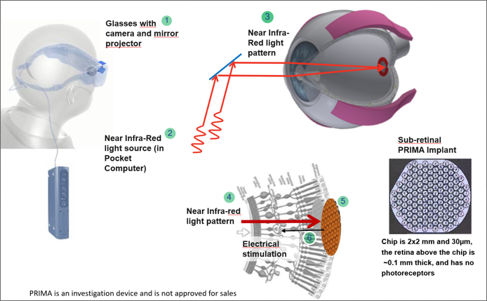
The patients also had to wear clear glasses fitted with a camera that projected infrared light onto the implanted chip’s pixelated surface, which then transmitted electric signals to the brain via the optic nerve.
The restored vision is not normal color vision, Olmos de Koo clarified, but black-and-white prosthetic vision that, in the study, enabled most patients to read words.
"Aside from the injections to slow it down, we have nothing to offer right now in terms of replacement or restoring lost vision. And that's why this device, the chip, is such a ray of hope for this group of patients," she said.
For news organizations: Download broadcast-ready soundbites and related multimedia of Olmos de Koo discussing the implant and the study. The authors' conflict of interest statements are in the published paper, which will be provided to journalists upon request.
For details about UW Medicine, please see our About page.