Implant may help brain rewire after stroke
UW Medicine surgeons have implanted a device they hope will stimulate a stroke patient's brain to restore lost function.Media Contact: Susan Gregg - sghanson@uw.edu, 206-390-3226
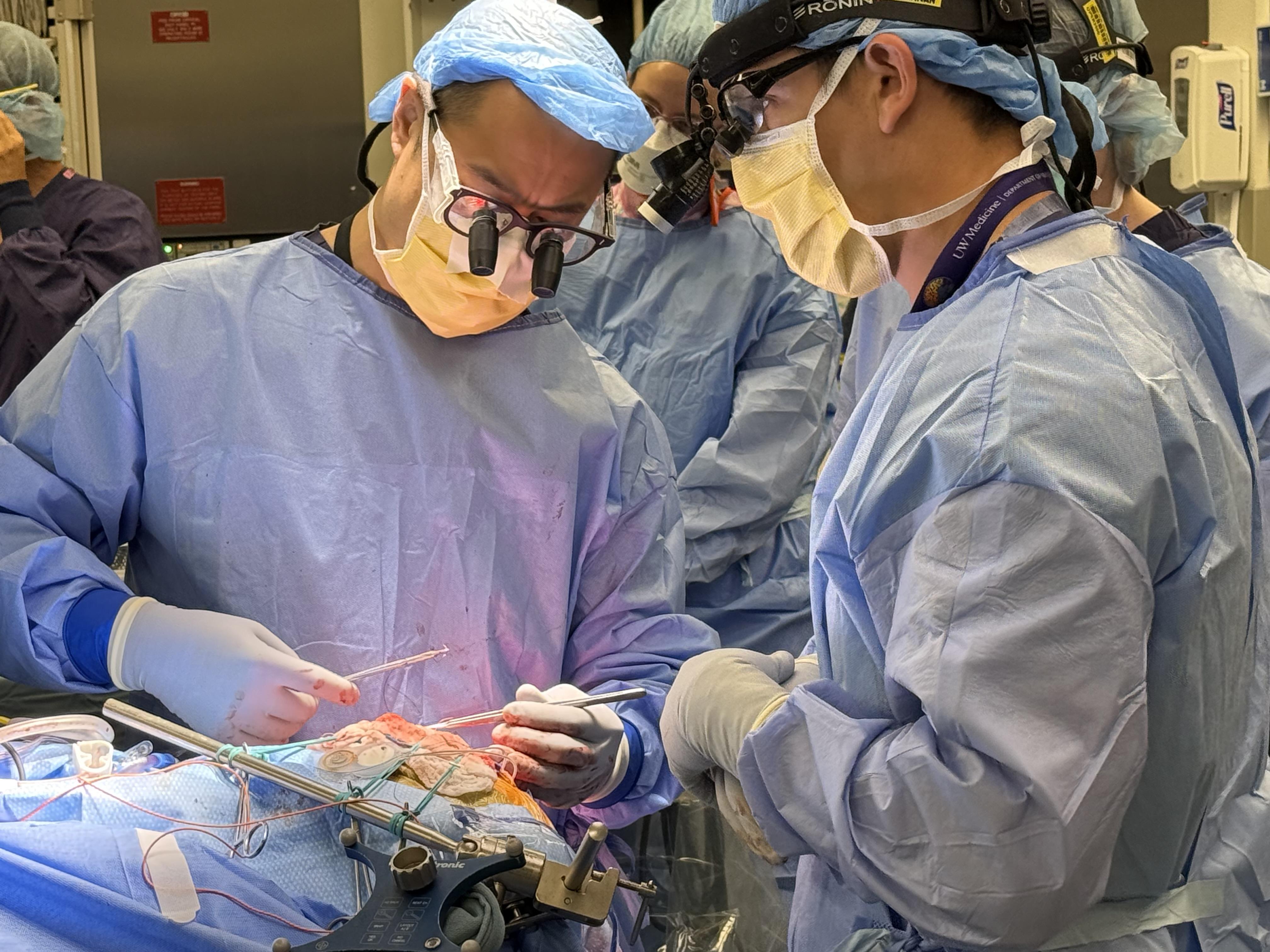
Neurosurgeons at the University of Washington School of Medicine have implanted a device they hope will help stroke patients recover lost function by stimulating their brains to rewire.
The device was implanted July 22 in a 52-year-old man who over the past years had several strokes that severely affected his ability to use his arm and leg. The patient recovered partial use of the limbs with rehabilitation, but the improvement was limited.
“People often get some function back after a stroke, but not all,” said neurosurgeon Jeffrey Ojemann, vice chair for discovery and professor of neurological surgery at the UW School of Medicine and the study’s co-principal investigator. “We want to see whether by stimulating the brain during rehabilitation sessions we can help them regain more function.”
Andrew Ko, an associate professor of neurological surgery at the UW School of Medicine, implanted the device in an operation at Harborview Medical Center in Seattle. He heads the UW Medicine Functional and Restorative Neurosurgery Program.
The study is being done in collaboration with the University of California, Los Angeles, where co-principal investigator and stroke neurologist Steven Cramer, UCLA professor of neurology, is leading the effort.
The project has a website with information about enrolling in the study.
The device is developed and manufactured by CorTec GmbH in Freiburg, Germany. This is the first time it has been used in a human. Its implantation, said CorTec CEO Frank Desiere, who holds a Ph.D. in life sciences, “marks a milestone for European medical neurotechnology and underscores CorTec’s emergence as Germany’s first implantable brain computer interface developer that is moving onto the global stage.”
Movement of the muscles — such as those that move the arms and legs — is controlled by a strip of interconnected neurons that runs over the top of the brain and is called the motor cortex. Thousands to tens of thousands of neurons may contribute to a neural circuit controlling a single movement.
These brain regions receive signals from many other parts of the brain and spinal cord. These regions then send the spinal cord signals that control the contraction of muscles. Some regions are primarily dedicated to the performance of a specific movement, whereas others may be dedicated to other contexts.
Strokes cause physical disability by destroying brain regions and connections involved in specific movements. Depending on the extent and location of the damage, the disability can be severe. In many cases, functional use of the affected body part (such as the hand) can be lost.
But if enough brain regions survive and remain connected, they can strengthen their existing connections and form new ones to help restore function, at least in part. In addition, brain regions that were not directly involved can be induced to contribute more to the circuit’s activity. Rehabilitation exercises help promote this rewiring by activating damaged circuits.
The study team is seeking to enlist this effect by using electrical impulses from the device to induce neurons to fire together when the patient tries to perform certain movements during rehabilitation sessions. The simultaneous firing of the neurons is thought to promote stronger connections between surviving regions.
Timing is crucial, said Ojemann “You want to activate the neurons when the brain is doing something you want to improve. The real power of this method is that it gets the two areas of the brain to train together.”
The implant consists of two soft, thin silicon sheets embedded with tiny electrodes. The sheets are placed on the surface of the brain over the area affected by the stroke. The electrodes can detect brain waves and emit electrical impulses to stimulate the brain.
The electrodes are connected to a small device implanted in the skull. This device wirelessly relays information through the skin to a computer that can analyze changes in brain waves and adjust the electrical impulses for maximum effect.
The study will include both rehabilitation sessions and extended monitoring of the patient’s neural signals, said Jeffrey Herron, an associate professor of neurological surgery at the UW School of Medicine who holds a Ph.D. in electrical engineering. He is an expert in brain-computer interfaces and will lead the study’s engineering aspects.
The device will be removed after about nine months.
“It’s a rehab enhancement device, not a treatment,” Herron said. “The idea is to implant the device, have the patient go through rehab while receiving stimulation, hopefully acquire long-lasting improvements, and then remove the device.”
Four patients will be enrolled in the safety trial. Another eight will be enrolled in a follow-up study.
The five-year study is funded by the National Institutes of Health’s National Institute of Neurological Disorders and Stroke (grant UH3NS121565).
written by Michael McCarthy
For details about UW Medicine, please see our About page.