Brain surgery patients help Allen Institute study
Cell samples, formerly discarded, are advancing science’s understanding of the brainMedia Contact: Susan Gregg - 206.390.3226, sghanson@uw.edu
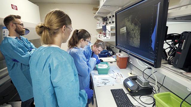
Researchers at the Allen Institute for Brain Science are using living cells from neurosurgery patients to explore and understand the brain’s building blocks. Patients at UW Medicine’s Harborview Medical Center and University of Washington Medical Center, as well as the Swedish Neuroscience Institute, are participating.
The tissue samples previously would have been discarded. Now they are being added to the Allen Cell Types Database, a publicly available tool.
“Our goal is to truly understand how the human brain works, not just the brains of mice or other animals that most brain scientists study. There is no better way to do that than to directly examine live cells of the human cortex,” said Christof Koch, chief scientist and president of the Allen Institute for Brain Science. “The human brain is the most complex piece of highly organized and active matter in the universe, and it is critical to understand its building blocks if we ever hope to comprehend how it works as a whole.”
Gathering this data is made possible through collaborations with Seattle-area neurosurgeons.
“Throughout the UW School of Medicine, we aim for a better understanding of the human body and human brain, and our collaboration with the Allen Institute has been a very natural one,” said Ojemann, professor of neurological surgery. “Their approach of thinking big and using high-throughput, large-scale methods has made them an exciting partner in our shared mission of understanding the brain. We will undoubtedly learn about many different diseases and disorders as we pursue this fundamental question of cell types in the brain.”
When patients require surgery for conditions like epilepsy or brain tumors, surgeons often need to remove a small piece of cortical tissue to reach the surgical target. This tissue is typically treated as medical waste and destroyed. Once transported to the lab, the tissue can be studied for a much longer time than tissue derived from the laboratory mouse, the most common animal model in the biomedical sciences.
“In the four years we have been conducting these experiments, we have found it is possible to work with the human brain tissue far longer than previously reported in the literature,” said Jonathan Ting, assistant investigator at the Allen Institute for Brain Science. “One of our hopes is that by pursuing work on living human brain cell types at a large scale, and by sharing our data publicly, we can inspire others in the scientific community to shift their own work toward human surgical tissue, in order to more directly address many of the scientific questions in the neuroscience field.”
By examining human brain cells in parallel to those of the mouse, this resource also allows comparison between species. While many general features of cellular structure and function are similar, the details can differ. For example, the shapes of human neurons are much larger and more complex, while the usage of many genes changes across species.
“We are beginning to find that certain types of neuronal and non-neuronal cells have very different properties in mouse and human,” said Ed Lein, investigator at the Allen Institute for Brain Science. “If we want to understand the specifics of human brain function in health and disease it is essential to study the human brain itself.”
The Allen Institute recently received nearly $100 million in funding from the National Institutes of Health, including nearly $34 million to lead a collaboration to further this work in studying and characterizing cell types in human tissue, and create a community resource for sharing the data.
The Allen Cell Types Database is freely available.
For details about UW Medicine, please see our About page.