3D-printed device advances human tissue modeling
The device is small enough to fit on a fingertip and can work with existing tissue-engineering technology.Media Contact: Leila Gray, 206-475-9809, leilag@uw.edu
A new, easily adopted, 3D-printed device will enable scientists to create models of human tissue with even greater control and complexity. An interdisciplinary group of researchers at UW Medicine and the University of Washington led the development of the device.
3D tissue engineering, which recently has undergone other major advances in speed and accuracy, helps biomedical researchers design and test therapies for a range of diseases.
One goal of tissue engineering is to create lab-made environments that recreate the natural habitats of cells.
Suspending cells in a gel between two freestanding posts is one of the current modeling platforms for growing heart, lung, skin and musculoskeletal tissues.
While this approach allows cells to behave as they would inside the body, it has not made it easy to study multiple tissue types together. More precise control over the composition and spatial arrangement of tissues would allow scientists to model complex diseases, such as neuromuscular disorders.
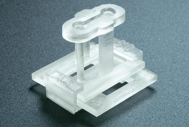
A paper published in Advanced Science details how the new platform lets scientists examine how cells respond to mechanical and physical cues, while creating distinct regions in a suspended tissue. The 3D-printed device is known as STOMP (Suspended Tissue Open Microfluidic Patterning).
Nate Sniadecki, professor of mechanical engineering and interim codirector of the UW Medicine Institute for Stem Cell and Regenerative Medicine, and Ashleigh Theberge, UW professor of chemistry, led the scientific team. The group showed that their device can recreate biological interfaces like bone and ligament, or fibrotic and healthy heart tissue.
The first authors of the paper were Amanda Haack, a student in the School of Medicine’s medical scientist program and postdoctoral fellow in the Theberge Lab, and Lauren Brown, a Ph.D. student in chemistry. UW faculty members Cole DeForest, professor of chemical engineering and bioengineering, and Tracy Popowics, professor of oral biology in the School of Dentistry, are coauthors.
STOMP enhances a tissue-engineering method called casting, which the researchers compared in simple terms to making Jell-O in a dessert mold. In the lab, the gel is a mixture of living and synthetic materials. These are pipetted into a frame rather than poured into a mold. STOMP uses capillary action — think of water flowing up a straw in a drinking glass — to permit scientists to space out different cell types in whatever pattern an experiment requires, like a cook evenly spreading pieces of fruit in Jell-O.
The researchers put STOMP to the test in two experiments: one that compared the contractile dynamics of diseased and healthy engineered heart tissue, and another that models the ligament that connects a tooth to its bone socket.
The STOMP device is about the size of a fingertip. It docks on to a two-post system originally developed by the Sniadecki Lab to measure the contractile force of heart cells. The tiny piece of hardware contains an open microfluidic channel with geometric features to manipulate the spacing and composition of different cell types, and for creating multiple regions within single suspended tissue without the need for additional equipment of capabilities.
Hydrogel technology from the DeForest Research Group souped up STOMP with another design feature: degradable walls. Tissue engineers can break down the sides of the device and leave the tissues intact.
“Normally when you put cells in a 3D gel,” Sniadecki said, “they will use their own contractile forces to pull everything together — which causes the tissue to shrink away from the walls of the mold. But not every cell is super strong, and not every biomaterial can get remodeled like that. So that kind of nonstick quality gave us more versatility.”
Theberge is excited about how other teams will use STOMP.
“This method opens new possibilities for tissue engineering and cell signaling research,” she said. “It was a true team effort of multiple groups working across disciplines.”
The National Institutes of Health (NIH) supported the research through R35GM128648, R35GM138036, R01HL149734, R03DE029827, T32CA080416, F30HL158030, R90DE023059 , 5TL1TR002318-08, and a supplement to R35GM128648. This work was also partially supported by the UW, Friends of FSH Research, The Chris Carrino Foundation for FSHD and fellowship funds from Senator Paul D. Wellstone Muscular Dystrophy Specialized Research Center – Seattle (NIAMS P50AR065139), and a gift from Ionis Pharmaceuticals.
Adapted from a feature article by Thatcher Heldring, UW Medicine Institute for Stem Cell and Regenerative Medicine.
For details about UW Medicine, please see our About page.