‘Sci-fi’ tech allows activation of brain cells in mice
Novel use of a powerful microscope might inform future therapies for substance-use disorders and anxiety.Media Contact: Chris Talbott - 206-543-7129, talbottc@uw.edu
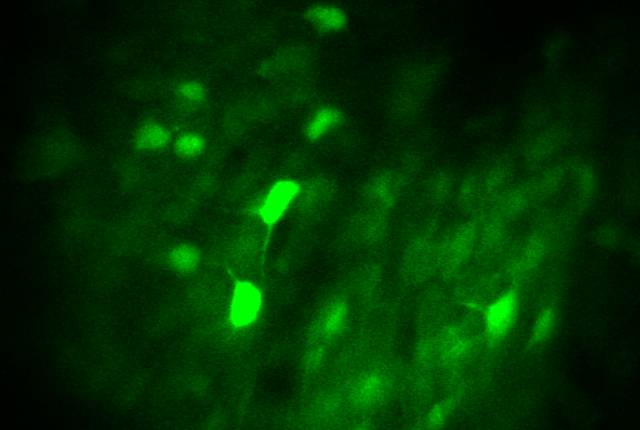
Using lasers and a holographic two-photon microscope that can peer deep into the brain, a team of scientists has identified and activated a small handful of cells among the hundreds of millions in a mouse's amygdala, a part of the brain that helps govern behavior.
The discoveries made in the lab of Michael R. Bruchas, professor of pharmacology and of anesthesiology and pain medicine at the University of Washington School of Medicine, might inform new treatments for substance-use disorders and an array of anxiety disorders.
“The technology is entering the realm of sci-fi,” said study co-author Zhe Charles Zhou, a research scientist and engineer who operated the microscope. “When I was a grad student 10 years ago, I never would have imagined this.”
While scientists have been able to stimulate groups of these intermingled neurons before, they haven’t been able to spatially separate them based solely on their observed activity. A two-photon microscope, with some of its components originally used in overhead projectors, allowed this group of researchers to not only watch reactions deep in the brain while a mouse ingested sweet or bitter solutions, but to stimulate the same very small groups of about a dozen neurons to alter behavior.
“We've been using light for over a decade to record and manipulate the activity of neurons,” said lead author Sean Piantadosi, a postdoctoral scholar in Bruchas’ lab. “But we’ve lacked the spatial precision to activate only specific ensembles within the deeper areas of the brain, in a physiological manner, and not (affect) nearby neurons that are not involved. With this technique, we can use holography to bend laser light to target solely the group of neurons that we want to manipulate.”
The work is highlighted in a Dec. 11 paper in Neuron. Researchers in the UW Department of Anesthesiology and Pain Medicine, the Center for the Neurobiology of Addiction, Pain and Emotion, the Department of Pharmacology and the Department of Bioengineering contributed to the investigation.
The team also showed stimulating one set of neurons inhibited rival neurons. They were surprised by the substantive reactions they got from such small groups of cells.
“I think one of the most interesting aspects our discovery, which gets to the core of neuroscience, is the question: What's the minimum number of neurons that might be regulating a given behavior?” Piantadosi said.
The almond-shaped amygdala is one of the most important regions in the brain, largely responsible for keeping humans safe from danger over millions of years of evolution. It is believed to govern fear and other emotional responses by helping to distinguish real-world threats such as dangerous animals from fake ones, for instance, in horror movies. But it also appears to have an underappreciated role in processing reward stimuli.
The two-photon microscope allowed the study’s authors to see the deep brain structure in a different light. Bruchas said there are indications that the amygdala plays a more nuanced role in feeling rewarded and in predicting and avoiding danger than previously thought.
Though this technique is unlikely to be repeated in humans, the group thinks the research could add to the understanding of how the brain governs decisions to consume or abstain from a substance.
“Thinking about the translational impact for human anxiety and substance-use disorders, our findings reveal a potential set of biological targets for future technology development aimed at mental health therapeutics,” Zhou said. “Finding a way to interact with the aversive cells Sean found, while minimizing negative side effects, could help across various mental health disorders, particularly drug addiction and anxiety disorders. That would be of one of our future goals.”
The Murdock Foundation provided funding for this study in conjunction with matched support from the University of Washington for the two-photon microscope used in this research. Additional support came from the National Institute of Mental Health (R01MH112355, R01MH111520), the National Institute on Drug Abuse (R37DA032750-10, F31DA051124, P30DA048736), the National Institute of General Medical Sciences (T32GM086270-12) and the Weill Neurohub.
For details about UW Medicine, please visit http://uwmedicine.org/about.