
CT scan radiation doses fell substantially in past decade
A radiology specialist credits advances in scanner technology, training, dose-monitoring, and more individualized patient parameters.Media Contact: Brian Donohue - 206-457-9182, bdonohue@uw.edu
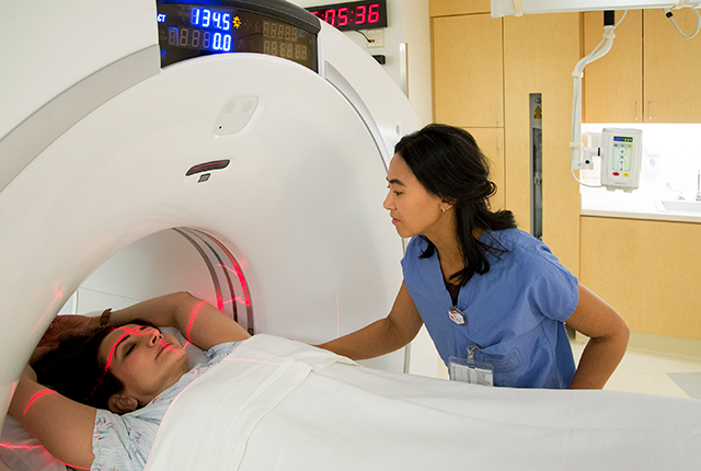
Radiation doses in common adult CT scans in the United States dropped substantially between 2014 and 2025, according to a study published in the journal Radiology.
CT, or computed tomography, uses ionizing radiation to generate cross-sectional images of internal organs and other tissues. These images are fundamental to helping doctors identify, monitor and treat hundreds of distinct medical conditions.
Over time, ionizing radiation can cause minor DNA damage that may convey a very small lifetime cancer risk. Cumulative exposure from multiple CT scans could increase that risk.
“Nationally, about 90 million CT scans are performed each year on about 60 million patients. So even small reductions in individual risk can have meaningful population-level health benefits,” said lead study author Kalpana Kanal, a medical physicist and professor of radiology at the University of Washington School of Medicine.
The study analyzed more than 5.2 million adult CT scans acquired in 2025 from 592 U.S. facilities. The sample was nearly four times larger than the comparison 2014 dataset. Across the 10 most common adult CT exam types, the levels of radiation decreased in two measures:
- Volume CT dose index: the actual dose output by a scanner during a given scan. A decrease of 21.8% was seen.
- Achievable dose: a benchmark that represents a dose level that can be achieved with optimized CT imaging protocols while maintaining the necessary image quality. A 9.3% decrease was seen.
Kanal said the lower doses reflect two decades of incremental gains: more sophisticated scanners, better training of clinicians, more precise CT scan parameters tailored to patient size, and accreditation programs’ validation of facilities’ good practice.
She also mentioned the use of artificial intelligence tools to further improve image quality: “This could have an impact on how we read scans that enables us to dial down the dose on the front end.”
Kanal emphasized that the link between ionizing radiation and cancer has been established primarily from studies of populations such as atomic bomb survivors, who were exposed to doses hundreds of times higher than those emitted in typical CT scans. Cancer risk estimates for CT scans and the general population have been extrapolated downward from those studies.
Here’s a more tangible reference point: A chest CT scan carries radiation roughly equal to two years of natural background radiation that U.S. residents receive from sources such as radon gas, soil and rock, and cosmic rays. A chest X-ray is roughly comparable to 10 days of background radiation.
Fears of developing cancer from CT scans should be weighed against the prospective upside of identifying a suspected health concern, Kanal said. In her view, “the benefit of getting a clinically indicated CT is much higher than the very small risk of having that radiation go into my body, even though it’s a carcinogen,” she said. “Undiagnosed conditions carry their own risk.”
Even so, she said patients who are referred for a CT scan should feel empowered to ask their physician what they expect to learn from it.
“We do a CT only if it is clinically indicated as the appropriate exam. Your doctor ought to be able to explain the potential benefit of a scan,” Kanal said.
For details about UW Medicine, please see our About page.