Project to map live-tissue molecular changes over time
Biohub funding will support creating a submicron-resolution platform to study immune dynamics in mouse and human tissues.Media Contact: Leila Gray - 206-475-9809, leilag@uw.edu
A two-year grant through Biohub’s Advancing Technologies for Spatiotemporal Omics in Live Tissue program will help a UW Medicine-led interdisciplinary research team develop a next-generation platform for molecular mapping in living tissues.
The team plans to apply their new technology to study immune dynamics in mouse and human tissues. The resulting longitudinal, multimodal datasets could also support AI-driven biomodels that predict changes in cellular behavior and tissue states over time.
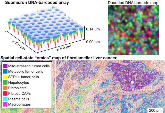
The project is a live-tissue spatiotemporal multi-omics platform centered on highly multiplexed proteomic measurements. Proteomics refers to the proteins being produced, modified or interacting in the tissue. This data will be integrated with gene-expression and immune-receptor readouts on the same tissue grid. The goal is to achieve submicron-scale spatial resolution. This is the ability to obtain measurements at dimensions of less than one micrometer, or 0.00004 of an inch.
The team aims to move beyond the static snapshots provided by fixed or frozen tissue methods and instead capture molecular changes as they unfold over time in living systems. This technology is being developed with the long-term goal of revealing how inflammation, immune responses, and disease processes evolve across space and time.
“Our goal is to make it possible to map molecular changes in living tissues as they happen, with exceptionally high spatial resolution,” said Liangcai Gu, associate professor of biochemistry at the University of Washington School of Medicine and coordinating principal investigator of the project. “That kind of dynamic, multimodal view could help us understand immune biology and disease progression in fundamentally new ways.”
The resulting longitudinal, multimodal datasets may also help support AI-driven biomodels that can forecast changes in cellular behavior and tissue states over time.
Biohub launched the program to accelerate minimally invasive, real-time profiling of molecular landscapes in live tissues, with a particular focus on immunology and autoimmune disease.
Collaborators on the project include Albert Folch, professor of bioengineering at the University of Washington; Zhicheng Ji, assistant professor of biostatistics and bioinformatics at Duke University; and Shreeram Akilesh, associate professor of laboratory medicine and pathology at the University of Washington School of Medicine. Folch and Ji are co-principal investigators on the project, and Akilesh is a co-investigator.
The project brings together expertise in chemistry, engineering, genomics, pathology, and computation. If successful, the platform could provide a broadly useful new technology for studying development, immunity, and disease with much richer spatial and temporal resolution.
The new effort builds on Pixel-seqV, a method the Gu lab played a significant role in pioneering. It uses DNA arrays called polony gels in mapping biomolecules and their interactions.
Related: Biohub blog item on grants awarded to develop technologies for spatiotemporal omics in living tissue,
For details about UW Medicine, please see our About page.