
Map reveals wiring in the human eye for best vision
The first census of neurons and their synapse-level connectivity in a fovea shows unique features of the human nervous system.Media Contact: Brian Donohue - 206-457-9182, bdonohue@uw.edu
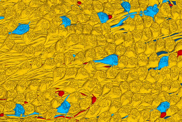
A new study provides an exquisitely detailed map of the circuitry of the fovea, the minute structure in the eye’s retina essential for seeing fine detail and discerning colors.
The map, called a connectome, describes the location of all the cells and connections between cells within the fovea, making it the first complete cell-type census and connectivity matrix of a human central nervous system structure.
"A connectome like this only recently became available for the fruit fly brain," said senior author Dennis M. Dacey. "The methods developed for the tiny fruit fly brain made it possible for us and other labs to attempt pieces of larger mammalian brains, and the transition to the human central nervous system was a real breakthrough for us and for understanding how human vision starts."
Dacey is a professor of neurobiology and biophysics at the University of Washington School of Medicine and a core scientist at the Washington National Biomedical Research Center. The study appears today in the Proceedings of the National Academy of Sciences.
The fovea is small, about the size of a pinhead, but it is packed with an enormous number of neurons, which enable detailed, high-resolution color vision. Although the fovea composes only a tiny portion of the retina, its signals account for the lion’s share of what reaches the brain’s visual centers.
It's the reason we swivel our eyes toward anything that catches our attention, whether it's a beautiful or terrifying sight. Pointing the fovea straight at something is the only way to see it in fine detail.
To build their map, the researchers reconstructed more than 300,000 connections among roughly 3,000 neurons in a human fovea, tracing exactly how each type of neuron links to all other neurons. They then compared this connectivity matrix to similar reconstructions from monkeys, which share a humanlike fovea, and mice, which lack a fovea entirely, as well as from the outer edges of the human retina, where vision is much less sharp.
The comparison turned up something striking: In most of the human retina, and in the other species, visual information travels through many different, overlapping circuits. But in the human fovea, evolution appears to have stripped things down to just three major pathways.
“Instead of many parallel routes, the fovea funnels information through a small number of highly efficient channels — something like a direct expressway rather than a tangle of side streets,” explained first author Yeon Jin Kim, research scientist in neurobiology and biophysics.
The researchers think this streamlined wiring may be what allows humans to support the demands of conscious, detailed vision and the complex behaviors that depend on it, such as reading a page or judging distance while driving. It may be a key piece of what sets human vision apart from that of other animals.
Such streamlining may have also occurred in other structures of the brain, perhaps explaining differences in human cognition, said Kim. “The retina is a part of the brain, so our analysis reveals what might be distinctive features of its neural circuitry. This map lets us start testing competing ideas about how those circuits compute.”
Beyond its implications for understanding the brain, the new connectome gives researchers a detailed blueprint of healthy foveal wiring. That blueprint could prove valuable for scientists working on treatments for blinding diseases that damage the fovea, since restoring vision will likely require rebuilding, or working around, this same intricate circuitry.
“The fovea is immensely important to human vision and quality of life," said co-author Christine Curcio, professor of ophthalmology at the University of Alabama at Birmingham. "The connectome is a tremendous resource for interpreting the cellular-level information available in diagnostic instruments used by ophthalmologists."
This work was supported by National Institutes of Health grants EY-028282, RF1-MH129260, P51-OD010425 and EY-01730.
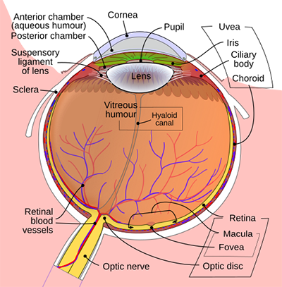
The image "Schematic diagram of the human eye with English annotations" was created by Rhcastilhos and Jmarchn and reprinted under the Creative Commons Attribution-Share Alike 3.0 Unported license.
For details about UW Medicine, please see our About page.